
Radiation oncology and image-guided assessment
We study image analysis for radiation oncology, including target segmentation and the interpretation of imaging changes after treatment.
How can image-derived boundaries and response measures be made more reproducible and interpretable?
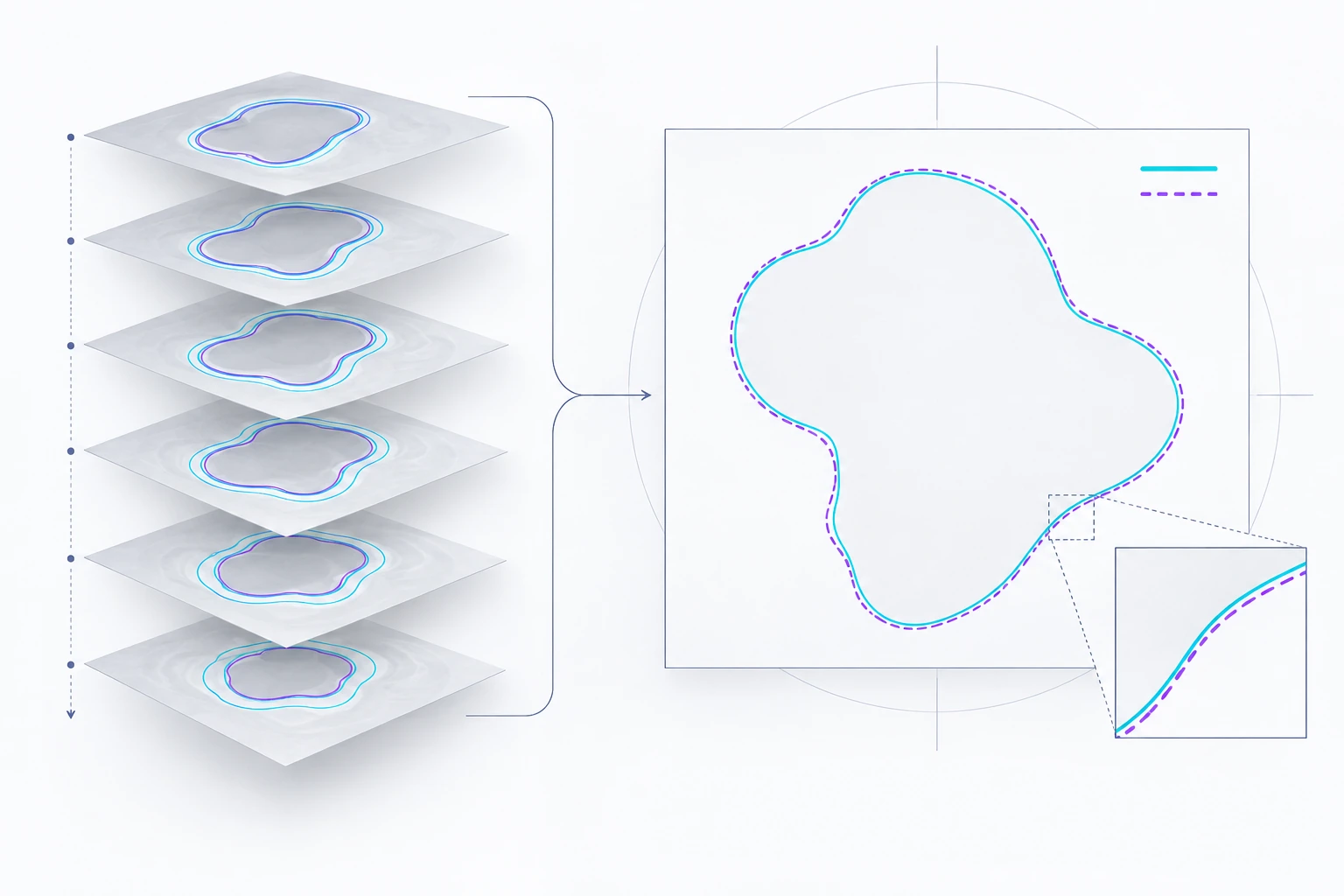
Method overview
Contours and imaging response
Reference contours specify the region to be analyzed across image slices.
Schematic of the research approach; image patterns and measurements are illustrative.
Research overview
Radiation oncology depends on carefully defined image regions and the interpretation of change over time. The lab’s research area includes FIDELIS and related work on automated image analysis. We examine how segmentation methods and imaging measurements can be evaluated against expert annotations and documented outcomes.
Representative collaborative work includes the BraTS-MEN-RT meningioma radiotherapy segmentation dataset. Other publications examine MRI response measures in soft-tissue sarcoma. These are distinct tasks: a contour describes an image region, while a response assessment asks what an observed change means. Their evaluation requires different reference standards.
Contours and imaging response
- Define annotated target regions and task-specific reference data.
- Compare model and expert contours, including their spatial disagreement.
- Investigate how imaging measurements relate to treatment-response reference outcomes.
Selected research sources
Research enquiries
Contact the lab