
Pancreatic imaging and cyst risk assessment
We develop image analysis methods to delineate pancreatic structures and investigate the imaging features associated with pancreatic cyst risk.
Which imaging representations support reproducible assessment of pancreatic cysts across institutions?
Method overview
Segmentation, representation, and risk
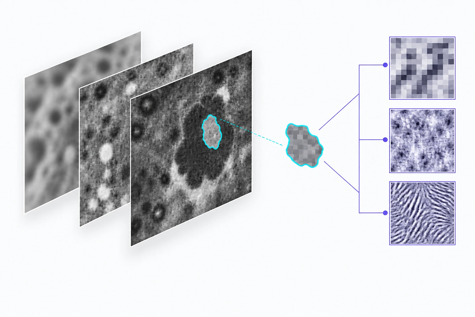
A region of interest separates the target from surrounding image information.
Schematic of the research approach; image patterns and measurements are illustrative.
Research overview
Pancreatic cyst assessment combines information about anatomy, image appearance, and clinical context. Our research studies segmentation, radiomic features, and learned representations from abdominal MRI. The aim is to characterize lesions consistently while examining how acquisition differences and limited labeled data affect model performance.
The Cyst-X research framework brings together a multi-center MRI benchmark, pancreatic segmentation, and malignancy-risk modeling. Related work on intraductal papillary mucinous neoplasms (IPMNs) compares centralized and federated training. These studies assess research models against documented reference outcomes; an imaging prediction is not itself a clinical diagnosis.
Segmentation, representation, and risk
- Delineate the pancreas and regions relevant to cyst assessment.
- Extract image features at multiple scales and combine complementary MRI information.
- Evaluate risk models across sites and compare approaches to collaborative training.
Selected research sources
Research enquiries
Contact the lab