
Liver imaging and cirrhosis assessment
We study liver segmentation and MRI representations of cirrhosis, with attention to altered morphology and complementary imaging sequences.
How can models characterize liver disease when anatomy and image appearance vary substantially?
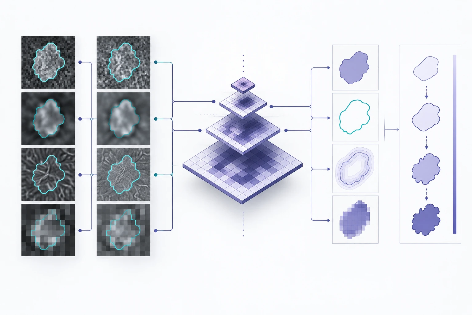
Method overview
Learning across MRI sequences
Contours define the liver region on complementary MRI sequences.
Schematic of the research approach; image patterns and measurements are illustrative.
Research overview
Cirrhosis changes liver morphology and image appearance, making automated analysis more difficult than delineating a healthy organ. Our work develops and evaluates segmentation methods on MRI with expert annotations, and investigates representations that distinguish disease stages.
CirrMRI600+ provides a research collection of T1- and T2-weighted MRI with cirrhotic liver segmentations. Our stage-estimation work combines multi-scale image information with attention across MRI sequences. Together, these studies examine both the definition of the imaging region and the features used to characterize it.
Learning across MRI sequences
- Define expert-annotated liver regions in heterogeneous MRI data.
- Learn features at several spatial scales from T1- and T2-weighted images.
- Assess segmentation and stage-estimation performance using the study’s reference labels.
Selected research sources
Research enquiries
Contact the lab