
Thoracic imaging and pulmonary disease
We investigate quantitative CT analysis of pulmonary disease, including imaging patterns associated with fibrosis after COVID-19.
How can image features describe pulmonary abnormalities and their relationship to subsequent disease?
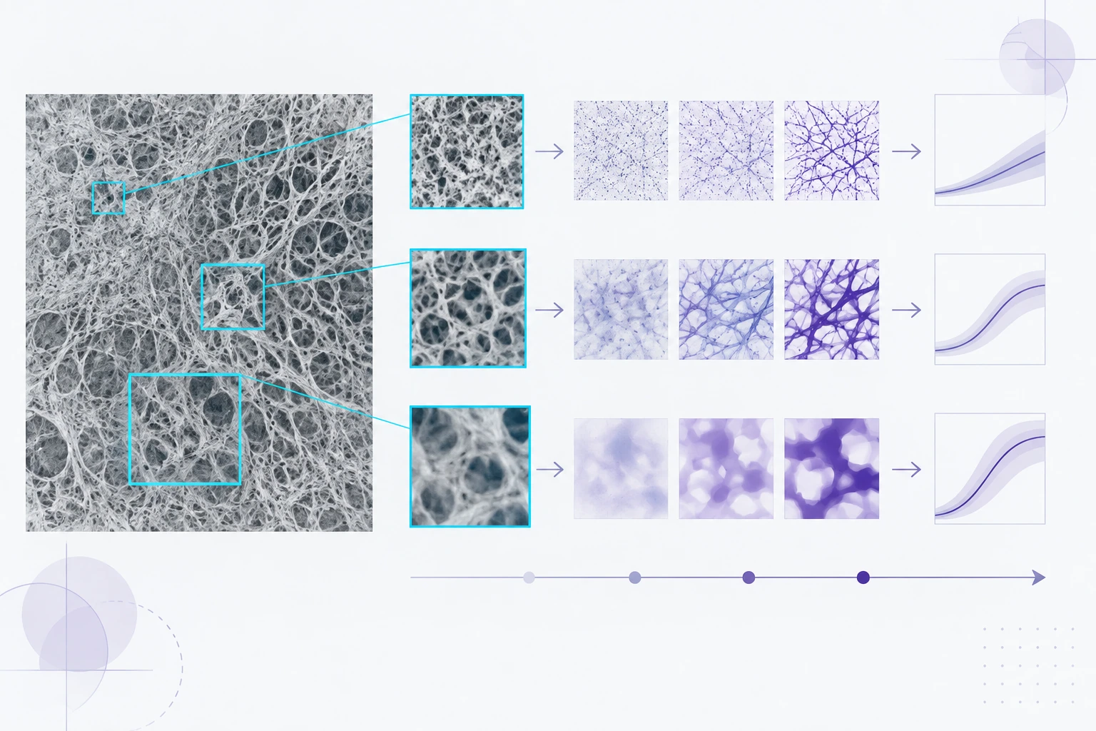
Method overview
Quantifying pulmonary image patterns
Image regions capture local appearance at different spatial scales.
Schematic of the research approach; image patterns and measurements are illustrative.
Research overview
Thoracic images contain patterns at several spatial scales, from local texture to the distribution of abnormalities across the lungs. Our research combines radiomics and deep learning to study these patterns in clinical imaging datasets. A central methodological issue is whether a model captures disease-related information that remains useful across patient groups and acquisition settings.
Our work on post-acute sequelae of COVID-19 (PASC) investigates CT-based prediction of pulmonary fibrosis formation. It compares image representations and examines regions contributing to model predictions. This sits within the lab’s broader research in lung imaging, disease characterization, and interpretable medical image analysis.
Quantifying pulmonary image patterns
- Identify image regions and patterns relevant to the pulmonary task.
- Compare radiomic texture descriptors with learned multi-scale features.
- Evaluate outcome prediction and inspect the image evidence associated with it.
Selected research sources
Research enquiries
Contact the lab